BASAL ULTRASONOGRAPHY
BASAL ULTRASONOGRAPHY
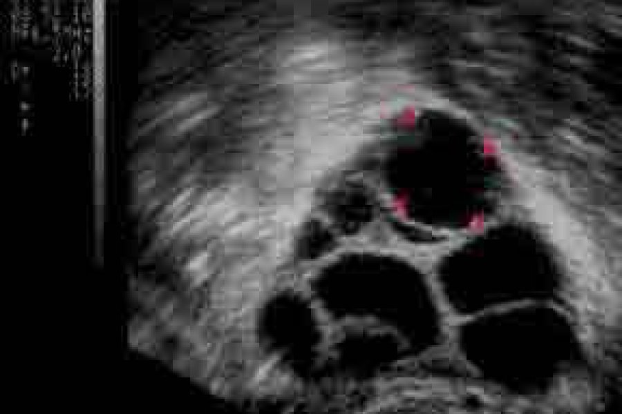
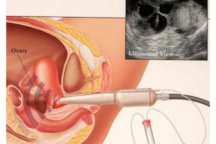
Ideally, it should be done on the 2nd or 3rd day of menstruation. Preferably, a vaginal probe is used. The uterus, ovaries, tubes, and Douglas cavity (the area behind the uterus) are evaluated. The size of the uterus, whether it contains fibroids, the presence of irregularities or polyps in the endometrium (inner layer of the uterus), and formations that press on the endometrium are evaluated. The sizes of both ovaries are measured separately, the antral follicles (egg precursors) in the ovaries are counted. Pathological formations that may be in the ovary (endometrioma (chocolate cyst) or ovarian cyst) are detected. It is checked whether there is a hydrosalfenx (the liquid-filled cystic and dysfunctional state of the tubes) in the tubes.
Pathologies detected by ultrasonography and reducing the success of IVF should be corrected before the procedure. For example, if there is a hydrosalfenx, it should be removed by laparoscopic surgery, if there is an endometrial polyp, it should be removed by hysteroscopy, if there is a fibroid pressing on the endometrium, it should be removed by surgery. Endometriomas (chocolate cysts) and ovarian cysts are approached according to the patient's condition.
